Article

Success and Complication Rates of Transvenous Lead Extraction in a Developing High-Volume Extraction Center: The Zurich Experience

Division of Electrophysiology, Department of Cardiology, University Heart Center, University Hospital Zurich, 8091 Zurich, Switzerland
*
Author to whom correspondence should be addressed.
J. Clin. Med. 2023, 12(6), 2260; https://doi.org/10.3390/jcm12062260
Submission received: 12 February 2023 / Revised: 6 March 2023 / Accepted: 8 March 2023 / Published: 14 March 2023
(This article belongs to the Section Cardiology)

Abstract

:
Introduction: Transvenous lead extractions are increasingly performed for malfunction or infection of cardiac implantable electronic devices, but they harvest a potential for complications and suboptimal success. Apart from multicenter registries and reports from highly experienced single centers, the outcome in individual newly developing high-volume centers starting a lead extraction program is less well established. We aimed to evaluate the clinical and radiological success and complication rate at our center, having started a lead extraction program less than a decade ago. Methods: We retrospectively analyzed patients who underwent transvenous lead extraction at the University Hospital Zurich from 2013 to 2021 regarding success as well as complications and compared our results to previously reported outcome rates. Results: A total of 346 patients underwent 350 transvenous lead extractions from January 2013 to December 2021. Combined radiological success was achieved in 97.7% and clinical success in 96.0% of interventions. Procedure-related major complications occurred in 13 patients (3.7%). Death within 30 days after transvenous lead extractions occurred in 13 patients (3.7%), with a procedure-related mortality of 1.4% (five patients). Summary: Transvenous lead extractions in newly developing high-volume centers can be performed with high clinical and radiological success rates, but procedure-related major complications may affect a relevant number of patients. Compared to large single or multicenter registries of experienced centers, the success rate may be lower and the complication rate higher in centers newly starting with lead extraction, which may have important implications for patient selection, procedural planning, proctoring, and safety measures.

1. Introduction

Since the introduction of transvenous cardiac pacing in the past century, the number of implantations of cardiac implantable electronic devices (CIED) has dramatically increased over the years, with by now approximately 1,000,000 de novo implanted transvenous leads every year worldwide [1]. The increase in implantation can largely be explained by population growth, longer life expectancy, facilitated healthcare access, and expanding indications for device therapy [2]. Because of lead dysfunctions or device infections, the number of transvenous lead extractions (TLEs) performed has also increased, with an estimated 10,000–15,000 device leads being extracted every year worldwide using specialized tools [3]. TLE techniques have been steadily improved to increase efficiency and safety, but the population in need of TLE is also becoming older and often demonstrates more comorbidities, more complex device systems, and more prior device interventions [4]. Previously available outcome rates of TLE are mainly derived from large single or multicenter registries of highly experienced extraction centers with a reported clinical success rate of 96.7 to 97.7, an overall combined radiological success rate of 93 to 99.3%, and a reported major procedure-related complication rate of 0.4 to 2.3% [5,6,7,8,9]. However, the outcome after TLE may differ in the real-world setting outside of highly experienced centers and the associated registries, with limited experienced operators, and in only just developing high-volume centers [10,11]. Our center started with a supervised and structured lead extraction program in 2013 and surpassed 30 TLEs per year as a standard for a high-volume center in 2018 with 51 TLEs, resulting in 72 TLEs in 2021 [7,12]. For this study, we retrospectively analyzed the success and complication rates of TLE in our developing high-volume center in the last 8 years, and compared the outcome to larger registries of centers with longer high-volume experience.

2. Materials and Methods

2.1. Study Design

We retrospectively analyzed all patients undergoing TLE at the university hospital Zurich from the beginning of 2013 (reflecting the start of a standardized and supervised extraction program) until the end of 2021. Every TLE procedure was considered as a separate entity, without regard to patient identity, in order to allow for multiple TLE within the same patient to be included in this study, as long as the second TLE procedure was not performed during the same hospital stay. Patients undergoing lead explantation (see Section 2.4 Definitions) were purposely excluded, as were patients undergoing lead extraction during open chest surgical approaches. Because of the retrospective design, the choice of extraction tools and their size was always left at the discretion of the operator. This study was approved by the local ethics committee (BASEC-NR: 2018-01540).

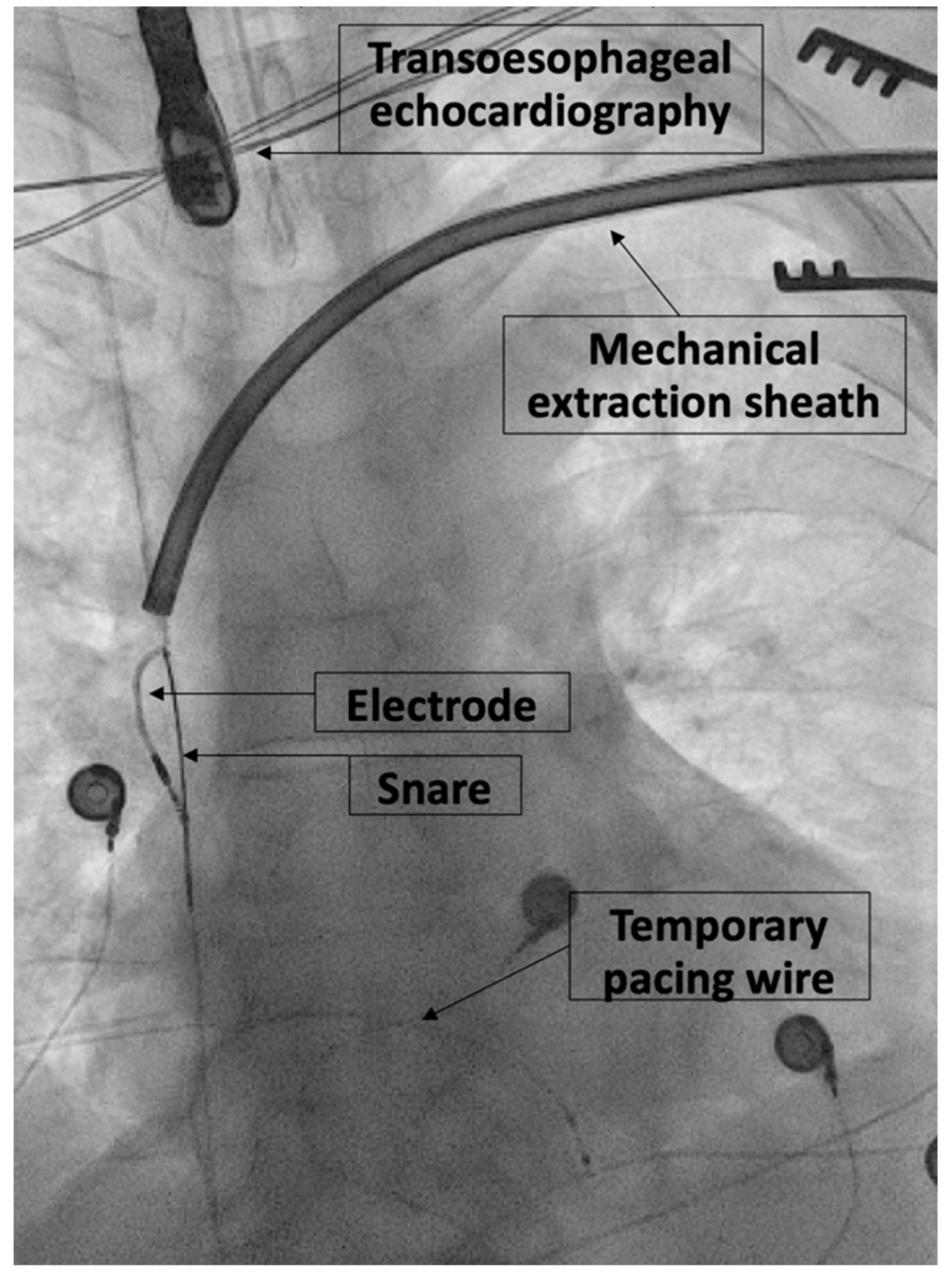
2.2. Extraction Procedure

Participating physicians were a cardiac surgeon at the beginning of the study period and three cardiologists with electrophysiology (EP) specialty in the latter part of the study period. TLE was performed under general anesthesia or conscious sedation based on the decision of the operator. TLE was performed in a hybrid operating room in the majority of procedures, and a standard electrophysiology laboratory in a minority of procedures. A superior approach via the implant-related vein was always the primary TLE method unless this approach had previously failed. If simple traction failed, a locking stylet (Liberator® Beacon® Tip Locking Stylet, Cook Medical, Bloomington, IN, USA) was introduced into the lumen of the lead and deployed. To increase lead control, a compression coil (One-Tie® Compression Coil, Cook Medical) was used. Because of historical extensive in-house experience with mechanical rotational lead extraction tools and concerns about safety with laser extraction tools, only mechanical rotational lead extraction tools (TightRail™ and TightRail Sub-C™ by Philips, Amsterdam, The Netherlands, or Evolution® RL and Evolution® Shortie RL by Cook Medical) were used during the study period. If the superior approach failed or additional stability during mechanical lead extraction was needed, a femoral approach using snares (i.e., Needle’s Eye Snare®, Cook Medical, or Amplatzer Goose Neck™, Medtronic, Dublin, Ireland) was performed. Some leads were removed using a combination of superior and femoral extraction approaches, especially if insufficient stability or traction during mechanical TLE was suspected (Figure 1).

2.3. Data Collection and Analysis

Patient cases were each individually assessed for evaluation of success, failure, and complications. Data collection was performed by studying operation reports, discharge reports, and chest X-rays. Continuous variables are expressed as mean ± standard deviation, while categorical variables are expressed as absolute numbers and percentages. All patient data were anonymized.

2.4. Definitions

Definitions were aimed to be as equivalent as possible to the 2009 and 2017 HRS expert consensus and the 2018 EHRA expert consensus statement on lead extraction as well as similar to definitions used in the large multicenter registries to allow comparison (Table 1) [7,12,13,14]. However, due to incomplete annotation in patient reports, some alterations were made in comparison with these expert consensus statements and large multicenter registries: Firstly, we did not differentiate between post-procedural or intra-procedural complications because of prominent lack of data availability in patient charts. Secondly, because ascription of complications may be challenging as patients may undergo additional procedures during their hospital stay, we decided to report all complications identified in medical charts during the hospital stay for TLE. Similar to the EHRA and HRS expert consensus statements, clinical success of TLE was possible in the absence of radiological success, because clinical success and failure of TLE mainly depend on the indication of TLE. In case of infection, only complete CIED removal should be considered a clinical success. In case of indications other than infection, clinical success may be granted despite a remnant of <4 cm, if the remaining lead presumably does not increase the risk of complications (i.e., vascular obstruction) and in the absence of procedure-related major complications [13]. Death after TLE was reported for up to 30 days after TLE in this study. Hereinafter we present our definitions for this study concerning the TLE procedure, success, failure, and complications.

3. Results

A total of 346 patients underwent transvenous extraction of 658 leads during 350 interventions from January 2013 to December 2021 and a mean follow-up time of 131.7 months. In four patients, TLE was performed twice during the study period (three for recurring lead defects and one for additional extraction with a jugular snare approach). TLE was performed by four operators during the study period, with one single operator performing 71.4% of all procedures. The mean age at the time of TLE was 63.6 ± 15.3 years, 71.7% of the patients were male, and the mean left ventricular ejection fraction (LVEF) was 45.7 ± 15.1% (Table 2). The main indication for TLE was lead dysfunction (50.3%), followed by infection (30.9%) and device upgrades (10.6%). The mean number of leads extracted per intervention was 1.9 ± 0.8, and the mean lead indwelling time was 112.5 ± 78.6 months (longest 392 months and shortest 13 months).

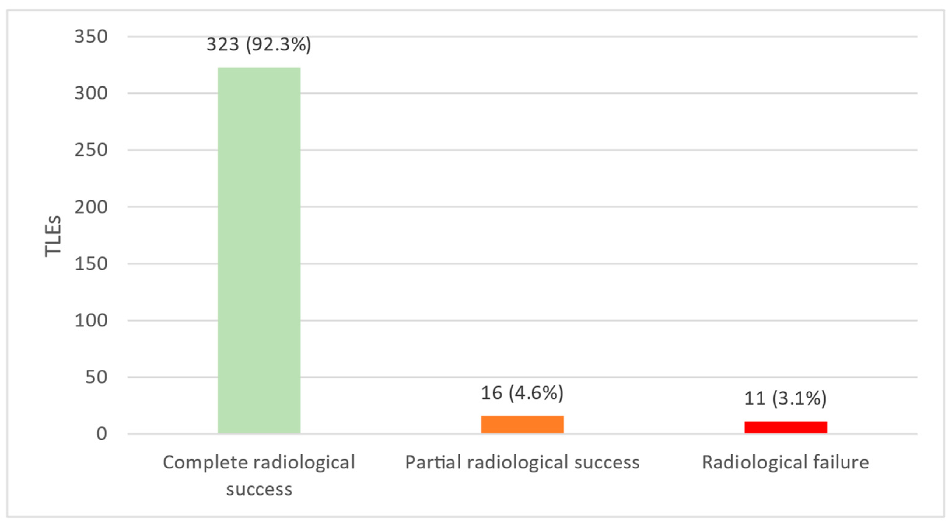
3.1. Radiological and Clinical Success
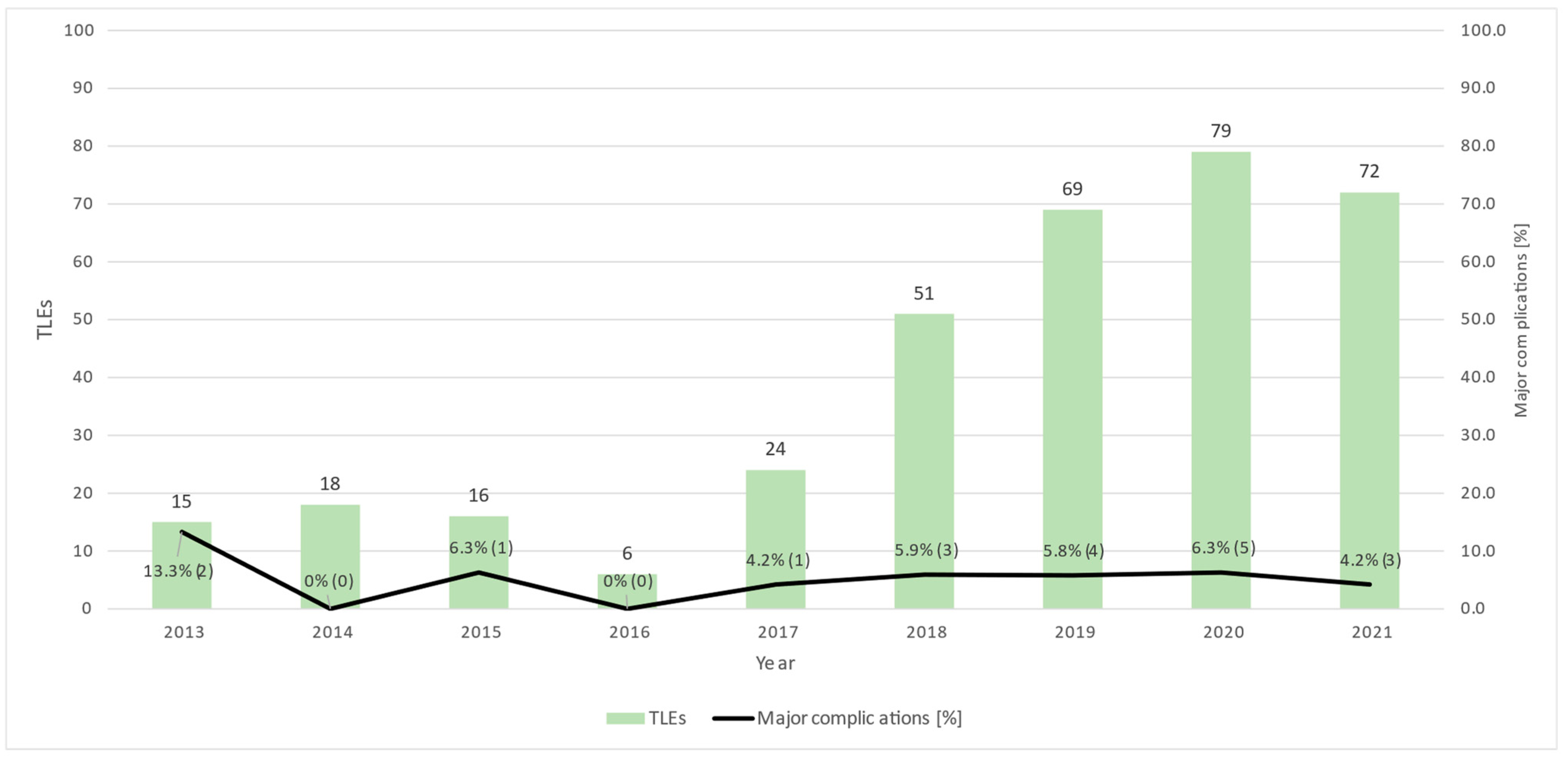
A total of 323 out of 350 TLE (92.3%) resulted in complete radiological success, and 16 TLE (4.6%) resulted in partial radiological success, cumulating in 96.8% of all TLE, resulting in combined radiological success. A number of 11 TLE procedures (3.1%) resulted in radiological failure (Figure 2). Clinical success was achieved in 329 out of 350 TLEs (94.0%), while 21 TLEs (6.0%) resulted in clinical failure (death within 30 days in 8 TLE, permanently disabling complications in 3 TLE, insufficiently extracted leads in 11 TLE, permanently disabling complications and insufficiently extracted leads in one patient).

3.2. Complications

Complications occurred in 78 of 350 TLEs (22.3%), of which 68 (19.4%) were minor, 19 (5.4%) were major, and a subgroup of 13 (3.7%) were procedure-related major complications (Figure 3). Reoperations due to complications had to be performed in 20 TLE (5.7%).

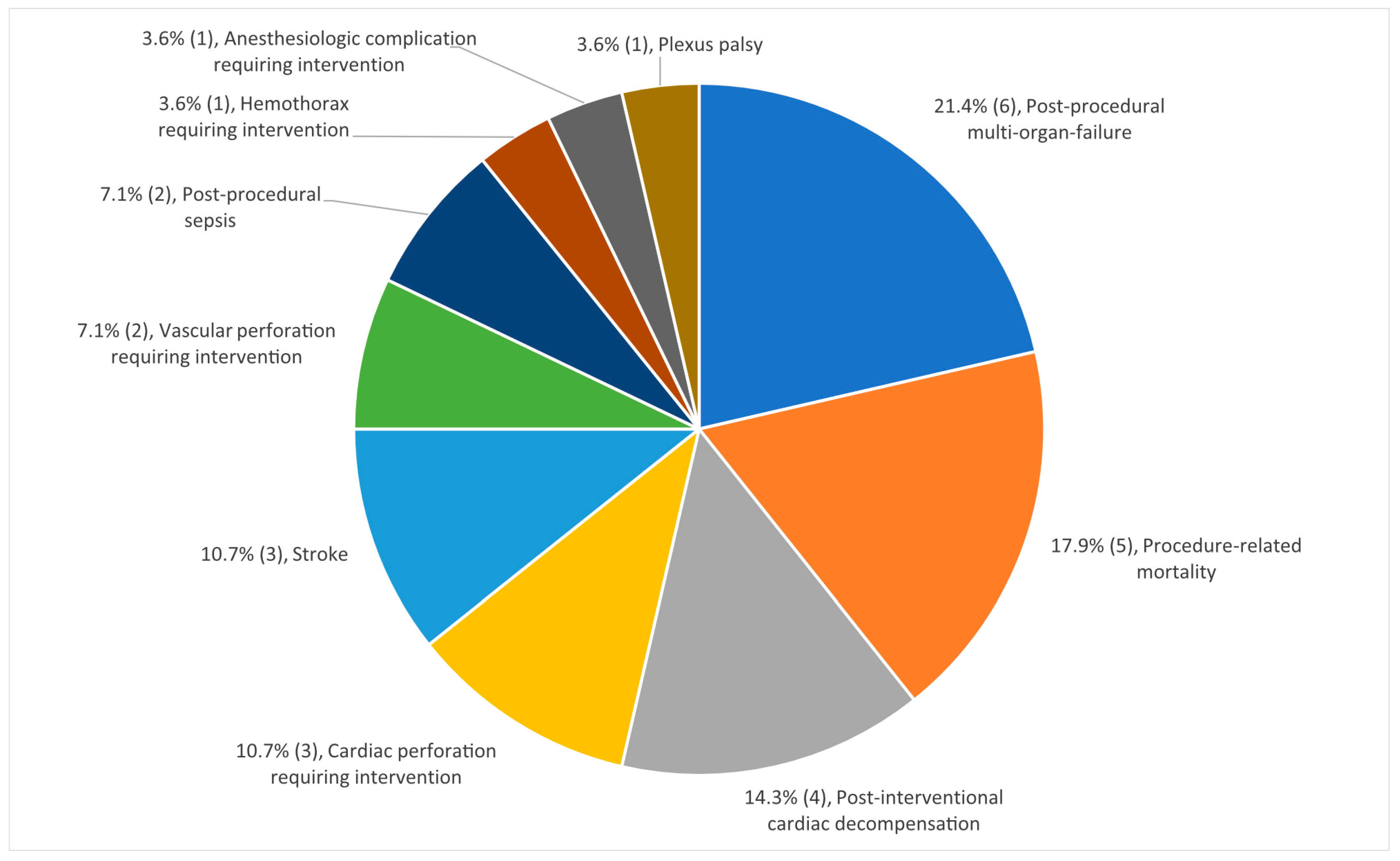
3.3. Major Complications

A total of 28 major complications occurred in 19 TLEs (5.4% of all 350 TLEs); the complications are divided into 14 different types (Table 3). The most common major complication was post-procedural multi-organ failure occurring after six TLEs (1.7% of all TLEs, 21.4% of all major complications): five patients developed multi-organ failure after TLE for infective endocarditis and concomitant surgical interventions and one patient developed multi-organ failure after hemorrhagic shock because of TLE-associated hemorrhage. Procedure-related mortality occurred after five TLEs (1.4% of all TLEs, 17.9% of all major complications). Post-interventional cardiac decompensation, cardiac perforation requiring interventions, and stroke followed thereafter (Figure 4). One patient required acute stenting of the vena cava superior for covered rupture of the vena cava superior during TLE, but no sternotomy had to be performed throughout the study period. While the amount of TLE/year increased over time, the yearly rate of major complications remained relatively stable at around 4-6% (Figure 5).

3.4. Procedure-Related Mortality

A total of 13 patients (3.7%) died within 30 days after TLE. However, only five deaths were related to the TLE procedure, resulting in a procedure-related mortality of 1.4% (Table 4). Another six patients died before discharge, and two patients passed away outside of the hospital within 30 days (in-hospital mortality of 3.1%). Out of the five presumably procedure-related deaths, two patients died due to a stroke, two patients because of post-interventional cardiogenic shock, and one patient due to post-interventional embolic distal aortic occlusion.

3.5. Minor Complications

A total of 82 minor complications occurred in 68 of 350 TLEs (19.4%). The most common minor complication was hematoma without any necessity of intervention (5.1%), followed by newly developed severe tricuspid regurgitation and swelling of the arm/thrombosis (each 2.9%), pericardial effusion without the need for intervention and blood transfusion (each 2.3%), and others (Table 5).

4. Discussion

Our retrospective analysis from a developing high-volume TLE center demonstrated a combined radiological success rate of 96.9% (complete and partial radiological success) and a complete radiological success rate of 92.3%. In comparison, the recent large single or multicenter registries demonstrated slightly higher rates of combined radiological success rate ranging from 98.5 to 99.3%, and complete radiological success ranging from 94.8 to 96.5% [5,6,7,15,16,17]. While there are also studies with lower radiological success rates, different definitions of success and failure complicate comparison [8,16]. Similarly, our clinical success rate of 94.0% was lower than reported success rates from larger established high-volume centers with 96.7–98.7% [5,7,15,16,17]. Regarding complications, we found a slightly higher rate of procedure-related major complications (3.7%) and a higher rate of minor complications (19.4%) compared to reported rates from most large registries (procedure-related major complications 0.4–2.6%, minor complications 1.4–7.2%) [5,6,7,8,9,15,16,17,18,19]. While there are also studies with a higher rate of procedure-related major complications, these often concern specific leads with known increased extraction difficulty [20]. Our rates of procedure-related (1.4%) and in-hospital mortality (3.1%) also appear to be slightly higher than reported mortality rates of experienced and established high-volume centers [5,7,15,17]. For specific complications, we also found post-procedural multi-organ failure and post-interventional decompensation as prominent major complications as opposed to primarily cardiovascular lesions in previous studies, while hematoma after TLE remained the most prominent minor complication, similar to previous studies [5,7,19].
In summary, our rates of radiological and clinical success were lower, and complication or mortality rates were higher than previously reported from experienced and established high-volume centers. We only reached the definition of a “high volume TLE center” with more than 30 TLE/year in the last 4 years of this study, and lower TLE experience has previously been shown to be a predictor of mortality and, therefore, procedure failure in TLE, so this may partially explain our results [21]. However, the rate of major complications per year remained relatively stable throughout the study period (Figure 5). Apart from less experience, differences in the definition of success and failure as well as patient and lead characteristics may provide alternative explanations for these differences: while mean patient age was similar in most studies (range from 62.866 years [5,7,15,17,18,19]), our patient population seemed to suffer more frequently from renal insufficiency (34.6% versus 17.4% [7]), potentially suggesting a population with more comorbidities overall. However, comorbidities and medication are sparsely represented in most registries [6,7,18], impeding coherent comparisons with our patient population. Concerning lead characteristics, the number of leads extracted per intervention, presence of ICD leads, and ratio of single/dual-coil ICD leads were similar to previous reports [5,6,7,15,18]. However, our leads demonstrated a definitely longer mean indwelling time of 9.4 years (112.5 ± 78.6 months) compared to the patient population of most previous larger registries with a range of 5.7 to 6.8 years [5,6,7,8,15,18,19]. This may be an important difference in the interpretation of our results, since longer lead dwelling time is a predictor of clinical and radiological failure, as well as complications [5,8]. Additionally, methods concerning the definition of complications and the timeframe of annotated complications vary between registries and publications [5,6,7,8,9,15,16,17,18,19]; we reported all complications during the hospital stay without adjudication in relation to the TLE procedure, which may also partially explain our higher rate of major and minor complications compared to previous publications.

5. Conclusions

Transvenous lead extractions can be performed with high clinical and radiological success rates, but procedure-related major complications may affect a relevant number of patients. Compared to large single or multicenter registries of experienced centers, the success rate may be lower and the complication rate higher in centers newly starting with lead extraction, which may have important implications for patient selection, procedural planning, proctoring, and safety measures.

6. Limitations of This Study

The single-center and retrospective design, sample size, and limited observation time are the main limitations of this study. Additionally, definitions of success, failure, and complication are heterogenous within previous studies, rendering comparison difficult. Another limitation of this retrospective analysis represents the missing data concerning individual success rates of TLE based on lead type, fixation, manufacturer, or location. Complications were only assessed within 30 days after TLE, rendering assumptions on long-term complications impossible.

Author Contributions

Conceptualization, D.H., J.S. and A.B.; methodology, D.H. and A.B.; validation, A.B.; formal analysis, D.H., N.K., M.C.B. and T.S.; investigation, D.H, T.S., J.S. and A.B.; resources, A.B.; data curation, D.H, N.K., M.C.B., T.S. and A.B.; writing—original draft, D.H. and N.K.; writing—review and editing, D.H., T.S., J.S. and A.B.; visualization, D.H. and A.B.; supervision, D.H., J.S. and A.B.; project administration, D.H. and A.B. All authors have read and agreed to the published version of the manuscript.

Funding

This research received no external funding.

Institutional Review Board Statement

This study was conducted according to the guidelines for good clinical practice and the Declaration of Helsinki. The gathering and analysis of all patient data for this paper adhere to the Swiss Ordinance on Human Research with the Exception of Clinical Trials (Human Research Ordinance and was approved by the local ethics committee (BASEC-NR: 2018-01540). All patient data were anonymized.

Informed Consent Statement

Informed consent was obtained from all subjects involved in the study.

Data Availability Statement

Upon urgent request and associated need, our data is available, while our utmost intention is to protect our patient’s privacy.

Conflicts of Interest

Daniel Hofer reports educational grants, consultant or speaker fees, and fellowship support from Abbott, Medtronic, Biotronik, Boston Scientific, Biosense Webster, Novartis, Bayer, Pfizer, and Spectranetics. Alexander Breitenstein has received consultant and/or speaker fees from Abbott, Bayer Healthcare, Biosense Webster, Biotronik, Boston Scientific, Bristol-Myers Squibb, Cook Medical, Daiichi Sankyo, Medtronic, Pfizer, and Spectranetics/Philipps. Jan Steffel has received consultant and/or speaker fees from Abbott, Alexion, Astra-Zeneca, Bayer, Berlin-Chemie, Biosense Webster, Biotronik, Boehringer-Ingelheim, Boston Scientific, Bristol-Myers Squibb, Daiichi Sankyo, Medscape, Medtronic, Menarini, Merck/MSD, Organon, Pfizer, Saja, Servier, and WebMD. He reports ownership of CorXL and Swiss EP. Steffel has received grant support through his former institution from Abbott, Bayer Healthcare, Biosense Webster, Biotronik, Boston Scientific, Daiichi Sankyo, and Medtronic. Sasse reports educational grants from Biotronik.

Abbreviations

TLETransvenous lead extraction
CIEDCardiac implantable electronic device
ICDImplantable cardioverter–defibrillator

References

  1. Diemberger, I.; Biffi, M.; Martignani, C.; Boriani, G. From lead management to implanted patient management: Indications to lead extraction in pacemaker and cardioverter-defibrillator systems. Expert Rev. Med. Devices 2011, 8, 235–255. Available online: https://pubmed.ncbi.nlm.nih.gov/21381913/ (accessed on 27 February 2023). [PubMed]
  2. Bongiorni, M.G.; di Cori, A.; Segreti, L.; Zucchelli, G.; Viani, S.; Paperini, L.; Menichetti, F.; Coluccia, G.; Soldati, E. Where is the future of cardiac lead extraction heading? Expert Rev Cardiovasc. 2016, 14, 1197–1203. Available online: https://pubmed.ncbi.nlm.nih.gov/27494362/ (accessed on 27 February 2023).
  3. Hauser, R.G.; Katsiyiannis, W.T.; Gornick, C.C.; Almquist, A.K.; Kallinen, L.M. Deaths and cardiovascular injuries due to device-assisted implantable cardioverter-defibrillator and pacemaker lead extraction. Europace 2010, 12, 395–401. Available online: https://pubmed.ncbi.nlm.nih.gov/19946113/ (accessed on 27 February 2023). [CrossRef] [PubMed]
  4. Kurtz, S.M.; Ochoa, J.A.; Lau, E.; Shkolnikov, Y.; Pavri, B.B.; Frisch, D.; Greenspon, A.J. Implantation trends and patient profiles for pacemakers and implantable cardioverter defibrillators in the United States: 1993–2006. Pacing Clin. Electrophysiol. 2010, 33, 705–711. Available online: https://pubmed.ncbi.nlm.nih.gov/20059714/ (accessed on 27 February 2023). [CrossRef] [PubMed]
  5. Wazni, O.; Epstein, L.M.; Carrillo, R.G.; Love, C.; Adler, S.W.; Riggio, D.W.; Shahzad, S.K.; Bashir, J.; Greenspon, A.J.; DiMarco, J.P.; et al. Lead extraction in the contemporary setting: The LExICon study: An observational retrospective study of consecutive laser lead extractions. J. Am. Coll. Cardiol. 2010, 55, 579–586. Available online: https://pubmed.ncbi.nlm.nih.gov/20152562/ (accessed on 27 February 2023). [CrossRef] [Green Version]
  6. Kennergren, C.; Bjurman, C.; Wiklund, R.; Gäbel, J. A single-centre experience of over one thousand lead extractions. Europace 2009, 11, 612. [Google Scholar] [PubMed]
  7. Bongiorni, M.G.; Kennergren, C.; Butter, C.; Deharo, J.C.; Kutarski, A.; Rinaldi, C.A.; Romano, S.L.; Maggioni, A.P.; Andarala, M.; Auricchio, A.; et al. The European Lead Extraction ConTRolled (ELECTRa) study: A European Heart Rhythm Association (EHRA) Registry of Transvenous Lead Extraction Outcomes. Eur. Heart J. 2017, 38, 2995–3005. Available online: https://pubmed.ncbi.nlm.nih.gov/28369414/ (accessed on 27 February 2023).
  8. Byrd, C.L.; Wilkoff, B.L.; Love, C.J.; Sellers, T.D.; Reiser, C. Clinical study of the laser sheath for lead extraction: The total experience in the United States. Pacing Clin. Electrophysiol. 2002, 25, 804–808. Available online: https://pubmed.ncbi.nlm.nih.gov/12049372/ (accessed on 27 February 2023).
  9. Sood, N.; Martin, D.T.; Lampert, R.; Curtis, J.P.; Parzynski, C.; Clancy, J. Incidence and Predictors of Perioperative Complications with Transvenous Lead Extractions: Real-World Experience with National Cardiovascular Data Registry. Circ. Arrhythm Electrophysiol. 2018, 11. Available online: https://pubmed.ncbi.nlm.nih.gov/29453324/ (accessed on 27 February 2023).
  10. Maytin, M.; Epstein, L.M. The challenges of transvenous lead extraction. Heart 2011, 97, 425–434. Available online: https://pubmed.ncbi.nlm.nih.gov/21296786/ (accessed on 27 February 2023). [CrossRef]
  11. di Monaco, A.; Pelargonio, G.; Narducci, M.L.; Manzoli, L.; Boccia, S.; Flacco, M.E.; Capasso, L.; Barone, L.; Perna, F.; Bencardino, G.; et al. Safety of transvenous lead extraction according to centre volume: A systematic review and meta-analysis. Europace 2014, 16, 1496–1507. Available online: https://pubmed.ncbi.nlm.nih.gov/24965015/ (accessed on 27 February 2023). [PubMed]
  12. Wilkoff, B.L.; Love, C.J.; Byrd, C.L.; Bongiorni, M.G.; Carrillo, R.G.; Crossley III, G.H.; Epstein, L.M.; Friedman, R.A.; Kennergren, C.E.H.; Mitkowski, P.; et al. Transvenous lead extraction: Heart Rhythm Society expert consensus on facilities, training, indications, and patient management: This document was endorsed by the American Heart Association (AHA). Heart Rhythm. 2009, 6, 1085–1104. Available online: https://pubmed.ncbi.nlm.nih.gov/19560098/ (accessed on 27 February 2023). [CrossRef] [PubMed]
  13. Bongiorni, M.G.; Burri, H.; Deharo, J.C.; Starck, C.; Kennergren, C.; Saghy, L.; Rao, A.; Tascini, C.; Lever, N.; Kutarski, A.; et al. 2018 EHRA expert consensus statement on lead extraction: Recommendations on definitions, endpoints, research trial design, and data collection requirements for clinical scientific studies and registries: Endorsed by APHRS/HRS/LAHRS. Europace 2018, 20, 1217. Available online: https://pubmed.ncbi.nlm.nih.gov/29566158/ (accessed on 27 February 2023). [PubMed]
  14. Kusumoto, F.M.; Schoenfeld, M.H.; Wilkoff, B.L.; Berul, C.I.; Birgersdotter-Green, U.M.; Carrillo, R.; Cha, Y.-M.; Clancy, J.; Deharo, J.-C.; Ellenbogen, K.A.; et al. 2017 HRS expert consensus statement on cardiovascular implantable electronic device lead management and extraction. Heart Rhythm 2017, 14, e503–e551. Available online: https://pubmed.ncbi.nlm.nih.gov/28919379/ (accessed on 27 February 2023). [CrossRef] [PubMed] [Green Version]
  15. Starck, C.T.; Gonzalez, E.; Al-Razzo, O.; Mazzone, P.; Delnoy, P.P.; Breitenstein, A.; Steffel, J.; Eulert-Grehn, J.; Lanmüller, P.; Melillo, F.; et al. Results of the Patient-Related Outcomes of Mechanical lead Extraction Techniques (PROMET) study: A multicentre retrospective study on advanced mechanical lead extraction techniques. EP Europace 2020, 22, 1103–1110. Available online: https://academic.oup.com/europace/article/22/7/1103/5843500 (accessed on 28 February 2023). [CrossRef] [PubMed]
  16. Migliore, F.; Pittorru, R.; Dall’Aglio, P.B.; de Lazzari, M.; Rovaris, G.; Piazzi, E.; Dentico, A.; Ferrieri, A.; D’Angelo, G.; Marzi, A.; et al. Outcomes of transvenous lead extraction of very old leads using bidirectional rotational mechanical sheaths: Results of a multicentre study. J. Cardiovasc. Electrophysiol. 2022. Available online: https://onlinelibrary.wiley.com/doi/full/10.1111/jce.15767 (accessed on 28 February 2023). [CrossRef]
  17. Sharma, S.; Lee, B.K.; Garg, A.; Peyton, R.; Schuler, B.T.; Mason, P.; Delnoy, P.P.; Gallagher, M.M.; Hariharan, R.; Schaerf, R.; et al. Performance and outcomes of transvenous rotational lead extraction: Results from a prospective, monitored, international clinical study. Heart Rhythm. O2 2021, 2, 113–121. Available online: https://pubmed.ncbi.nlm.nih.gov/34113913/ (accessed on 28 February 2023). [CrossRef]
  18. Jones IV, S.O.; Eckart, R.E.; Albert, C.M.; Epstein, L.M. Large, single-center, single-operator experience with transvenous lead extraction: Outcomes and changing indications. Heart Rhythm 2008, 5, 520–525. Available online: https://pubmed.ncbi.nlm.nih.gov/18325849/ (accessed on 27 February 2023). [CrossRef]
  19. Bongiorni, M.G.; Soldati, E.; Zucchelli, G.; di Cori, A.; Segreti, L.; de Lucia, R.; Solarino, G.; Balbarini, A.; Marzilli, M.; Mariani, M. Transvenous removal of pacing and implantable cardiac defibrillating leads using single sheath mechanical dilatation and multiple venous approaches: High success rate and safety in more than 2000 leads. Eur. Heart J. 2008, 29, 2886–2893. Available online: https://pubmed.ncbi.nlm.nih.gov/18948356/ (accessed on 27 February 2023). [CrossRef] [Green Version]
  20. Parkash, R.; Crystal, E.; Bashir, J.; Simpson, C.; Birnie, D.; Sterns, L.; Exner, D.; Thibault, B.; Connors, S.; Healey, J.S.; et al. Complications associated with revision of sprint fidelis leads: Report from the canadian heart rhythm society device advisory committee. Circulation 2010, 121, 2384–2387. Available online: https://www.ahajournals.org/doi/abs/10.1161/CIRCULATIONAHA.109.924357 (accessed on 27 February 2023).
  21. Sidhu, B.S.; Gould, J.; Bunce, C.; Elliott, M.; Mehta, V.; Kennergren, C.; Butter, C.; Deharo, J.-C.; Kutarski, A.; Maggioni, A.P.; et al. The effect of centre volume and procedure location on major complications and mortality from transvenous lead extraction: An ESC EHRA EORP European Lead Extraction ConTRolled ELECTRa registry subanalysis. EP Eur. 2020, 22, 1718–1728. Available online: https://academic.oup.com/europace/article/22/11/1718/5874070 (accessed on 1 March 2023). [CrossRef] [PubMed]
Figure 1. X-ray during lead extraction with combined superior and femoral approach.
Jcm 12 02260 g001
Figure 2. Radiological success or failure following transvenous lead extraction. Out of 350 TLE performed (100%), 323 TLE (92.3%) resulted in complete radiological success, 16 TLE (4.6%) resulted in partial radiological success, and 11 TLE (3.1%) resulted in radiological failure. TLE: transvenous lead extraction.
Jcm 12 02260 g002
Figure 3. Complications following transvenous lead extraction. Out of 350 TLE performed (100%), 272 TLEs (77.7%) went flawlessly. A total of 68 TLEs (19.4%) resulted in minor complications, 19 TLEs (5.4%) resulted in major complications, and a subgroup of 13 TLEs (3.7%) resulted in major procedure-related complications. One procedure may be listed in multiple groups. TLE: transvenous lead extraction.
Jcm 12 02260 g003
Figure 4. Frequency of major complications. Absolute number in relation to 350 TLEs in brackets. TLE: transvenous lead extraction.
Jcm 12 02260 g004
Figure 5. Ratio between TLEs performed and major complications by year. There was an overall increase in TLEs performed from 2013 to 2021. However, the absolute number of major complications was stable, with a subsequent decrease in relative risk. TLE: transvenous lead extraction.
Jcm 12 02260 g005
Table 1. Definitions. TLE: transvenous lead extraction.
Lead explantationRemoval of the lead by simple traction techniques and lead dwelling time below one year.
Lead extractionRemoval of at least one lead with a dwelling time of more than one year.
Complete radiological successRemoval of all targeted leads and material without any indwelling lead remaining.
Partial radiological successLead remnants <4 cm after TLE.
Radiological failureIncapacity to remove lead, >4 cm of targeted lead remaining.
Clinical successAchieving the clinical result for which the TLE was performed. Complete radiological success or partial radiological success, when the remaining lead fragment <4 cm, does not increase the risk of further complications, such as perforation, embolic events, conservation of infection, or any other undesired outcome. Absence of any procedure-related major complication resulting in permanent disability or death.
Clinical failureIncapacity to achieve clinical success.
Major complicationsAll complications causing persistent or significant disability, life-threatening events or death, or necessitating surgical intervention to prevent the above.
Procedure-related major complicationsComplications presumably related to TLE necessitating surgical intervention, requiring extension of hospitalization, causing persistent or notable disability, life-threatening events, or death.
Minor complicationsAny undesired event occurring during the same hospital stay as TLE requiring medical intervention, observation, or minor procedural intervention without requiring extension of hospitalization, causing persistent or notable disability, life-threatening events, or death.
ReoperationSurgical or interventional operation during the same hospital stay caused by a complication after TLE (procedure-related or unrelated).
Table 2. Patient, procedure, and lead characteristics.
Patients treated350
Mean age (years)63.6 ± 15.3
Male sex 251 (71.7%)
LVEF < 50%168 (48.0%)
LVEF absolute (%)45.7 ± 15.1
Hypertension 202 (57.7%)
Diabetes 73 (20.9%)
Renal insufficiency (eGFR < 60 mL/min) 121 (34.6%)
Anticoagulants
Single antiplatelet therapy
Dual antiplatelet therapy
Oral anticoagulation
Oral anticoagulation and antiplatelet therapy
None

79 (22.6%)
6 (1.7%)
148 (42.3%)
16 (4.6%)
101 (28.9%)
Indication for TLE
Lead dysfunction
Infection
Device upgrade
Other

176 (50.3%)
108 (30.9%)
37 (10.6%)
29 (8.3%)
Procedure average duration (min)131.7 ± 66.3
Mean lead dwell time (months)112.5 ± 78.6
Type of lead
Passive fixation
ICD leads
Dual-coil ICD leads
Right ventricular leads
Right atrial leads
Left ventricular leads (coronary sinus)

115 (32.9%)
158 (45.1%)
76 (21.7%)
194 (55.4%)
243 (69.4%)
69 (19.7%).
Mean number of leads extracted
Extracted leads per patient
1
2
3
4
5
1.9 ± 0.8

117 (33.4%)
165 (47.1%)
51 (14.6%)
12 (3.4%)
2 (0.6%)
Device reimplantation263 (75.1%)
Specific patient and lead characteristics of this study population. Other indications for TLE include chronic pain, abandoned or recalled leads, and other lead-related complications. LVEF: left ventricular ejection fraction. eGFR: estimated glomerular filtration rate. TLE: transvenous lead extraction. ICD: implantable cardioverter–defibrillator. Plus–minus values are means ± SD. Percentages in relation to procedures performed.
Table 3. Major complications.
Type of Major ComplicationNumber of Affected Patients
Post-procedural multi-organ failure6 (1.7%)
Procedure-related mortality5 (1.4%)
Post-interventional cardiac decompensation4 (1.1%)
Cardiac perforation requiring intervention3 (0.9%)
Stroke3 (0.9%)
Vascular perforation requiring intervention2 (0.6%)
Post-procedural sepsis2 (0.6%)
Hemothorax requiring intervention1 (0.3%)
Anesthesiologic complication requiring intervention1 (0.3%)
Plexus palsy1 (0.3%)
Type of major complication on the left, absolute number of those complications, and percent of all 350 TLE (=100%) in brackets on the right. TLE: transvenous lead extraction.
Table 4. Mortality after transvenous lead extraction.
Specific Mortality Number of Affected Patients
Procedure-related mortality5 (1.4%)
In-hospital mortality11 (3.1%)
All-cause mortality within 30 days13 (3.7%)
Specific subgroups of mortality after transvenous lead extraction. Percentages are calculated from 350 performed extractions (=100%).
Table 5. Minor complications after transvenous lead extraction.
Type of Minor ComplicationNumber of Affected Patients
Hematoma without need for intervention18 (5.1%)
Newly developed severe tricuspid regurgitation10 (2.9%)
Swelling of the arm/thrombosis10 (2.9%)
Pericardial effusion without need for intervention8 (2.3%)
Blood transfusion8 (2.3%)
Acute renal failure7 (2%)
Vascular complications with minor intervention6 (1.7%)
Hematoma with surgical revision5 (1.4%)
Electrode dislocation requiring revision4 (1.1%)
Pneumothorax requiring drainage2 (0.6%)
Pulmonary embolism2 (0.6%)
Anesthesiologic complication2 (0.6%)
Type of minor complication on the left, absolute number of those complications, and percent of all 350 TLE (=100%) in brackets on the right. TLE: transvenous lead extraction.
Disclaimer/Publisher’s Note: The statements, opinions and data contained in all publications are solely those of the individual author(s) and contributor(s) and not of MDPI and/or the editor(s). MDPI and/or the editor(s) disclaim responsibility for any injury to people or property resulting from any ideas, methods, instructions or products referred to in the content.

